Dongguan Aier Eye Hospital: One City, Three Hospitals and One Clinic, Part of a Global Network
 0769-22660023 0769-22660023
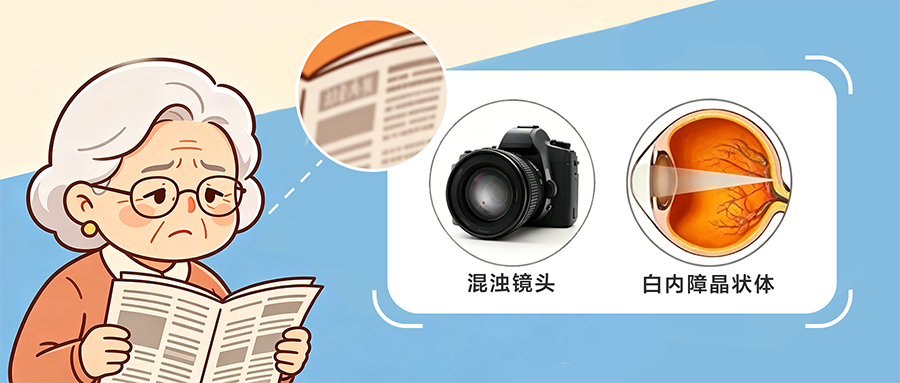
People often liken the eye to a precision camera, and this analogy is by no means an exaggeration. Our cornea acts as the camera’s outermost protective lens, the pupil is the aperture that regulates the amount of light entering, and the lens is the camera’s core—a natural zoom lens responsible for focusing and forming an image. The retina, located at the very back of the eye, serves as the light-sensitive film that captures the image.
The fundamental principle behind our ability to see clearly is as follows: light passes through the transparent cornea → through the pupil, which can expand or contract → through the clear, transparent and elastic lens → and is precisely focused onto the retina, allowing the brain to receive a clear and complete image.

A cataract, in essence, is when this natural lens becomes yellowed, cloudy, hardened and blurred.
A healthy lens: a ‘high-definition glass lens’
In youth, the lens is clear, transparent and elastic, much like a brand-new high-definition lens. It automatically thins when looking into the distance and thickens when looking at close objects, maximising light transmission. Light passes through unimpeded, allowing for clear vision without blurring or dark shadows.
As we age, everyone’s lens gradually deteriorates, much like the window panes in our homes that, after years of exposure to the elements, slowly oxidise, yellow and become dusty. Age-related cataracts are an inevitable part of the ageing process for everyone; it simply happens sooner for some and later for others.
How cataracts develop: the lens gradually ‘rusts and becomes cloudy’
Our lens is composed of layers of fibrous structures and undergoes constant metabolic processes throughout our lives. Under normal metabolic conditions, its internal structure is orderly, well-hydrated and uniform in texture. However, due to ageing, UV exposure, the natural ageing process and the accumulation of oxidative damage, the proteins within the lens undergo denaturation, coagulation and clumping. It is much like pouring milk into a glass of clear water, causing it to gradually become cloudy. At the same time, water is lost and the fibres harden; the originally transparent glass lens gradually transforms into frosted glass, sandblasted glass or yellowed, old glass.
Initially, there is only a slight haze at the edges of the lens, causing mild blurred vision; gradually, the cloudiness spreads from the edges to the centre, as if a thick fog has settled right in the middle of the lens; eventually, the entire lens becomes greyish-white, cloudy and opaque, preventing light from reaching the retina. This is advanced cataract, leading to near-total blindness.
These 3 groups of people experience lens deterioration earlier than others
People with High Myopia: ‘Thin, Fragile Lenses’ That Age Prematurely
In people with high myopia, the eyeball is elongated and stretched, much like a round balloon blown into an oval shape. As the eyeball is constantly stretched and expanded, the lens is also pulled and compressed, accelerating its ageing. Whilst others may develop cataracts at 60, those with high myopia may find them appearing as early as 40.
Furthermore, the lens capsule becomes thinner and less resilient, leaving the entire lens brittle and loose. Not only is it prone to clouding, but it is also often unstable in position and has a thinner posterior capsule. Cataracts progress much faster than in the general population, and surgery is more challenging.
To sum it up: whilst the average person’s lens is a durable model, the lens of a highly short-sighted person is a thin, brittle, and aged version subjected to prolonged strain—it fails sooner and deteriorates more rapidly.
Diabetes and Cataracts: A Lens ‘Damaged by a Sugar Solution’
Chronic hyperglycaemia is akin to soaking the eyeball in a corrosive sugar solution. Excess glucose in the blood seeps into the lens in large quantities, disrupting normal metabolic processes.
Sugar undergoes chemical reactions within the lens, producing a host of harmful substances that directly damage the lens proteins. The lens rapidly changes from a transparent state to becoming diffusely white and cloudy, much like clean glass left soaking in sugar water for a long time, gradually becoming mouldy, encrusted and veiled in a white haze.
Diabetic cataracts have two defining characteristics: they develop extremely rapidly and the clouding is exceptionally uniform. For many people with diabetes, vision can plummet dramatically within just a few months. Worse still, high blood sugar also damages the blood vessels at the back of the eye (akin to a ‘major traffic jam on the retinal motorway’), resulting in a situation where both the lens is damaged and the film is ruined—making it far more complex than a standard cataract.
Glaucoma Complicated by Cataracts: A ‘Clouded Lens’ Deformed by High Pressure
The core issue in glaucoma is persistently elevated intraocular pressure, leaving the eyeball in a state of constant high pressure and distension. Prolonged high pressure continuously compresses the lens, causing the internal fibres to become disorganised and accelerating protein denaturation and clouding.
Furthermore, the lens gradually swells and thickens, which in turn encroaches on the space within the eye, blocking the channels through which aqueous humour drains. This further elevates intraocular pressure, creating a vicious cycle where cataracts exacerbate glaucoma and glaucoma accelerates the progression of cataracts.
Such a lens is not only cloudy but also swells, thickens and bulges forward, resembling a frosted lens that has been squashed out of shape. It both blocks light and obstructs the flow of aqueous humour, making it particularly prone to triggering acute attacks of glaucoma.
How is cataract surgery performed? Why is an artificial lens implanted?
Where is the surgery performed? The entire procedure takes place in the anterior segment of the eye, at the edge of the cornea and in front of the pupil. It does not involve the back of the eye or the retina, so there is no need to worry about ‘going deep into the eye’. The surgeon makes a 2–3-millimetre micro-incision at the edge of the cornea (the limbus), which is about the size of a pinhead and does not require stitches.
Surgical Steps (In Plain English)
Step 1: The outermost transparent capsule of the lens—its protective shell—is opened.
Step 2: Using an ultrasound device, the natural lens inside, which has become cloudy, damaged or degenerated, is broken up and completely removed. This is akin to taking out a completely worn-out lens from a camera.
Step 3: The healthy lens capsule is retained; this capsule acts as a natural ‘retention slot’.
Step 4: Through the small incision, a high-definition intraocular lens is folded and inserted, securely locking into the capsule to replace the original natural lens.
Why can’t we just ‘clean it’—why must we replace the lens?
A cloudy lens is the result of complete protein denaturation and a total breakdown of its structure; it is not simply a case of a dusty surface. Neither medication nor eye drops can reverse this; the damaged lens must simply be replaced.
Without a lens, the eye is like a camera with its lens removed – it has absolutely no ability to focus. Even if the retina is perfectly healthy, vision will always be severely hyperopic and completely blurred.
An intraocular lens is essentially a bespoke replacement lens: it is made from high-tech medical-grade light-transmitting materials that remain permanently clear, do not age, and will not develop cataracts again. The surgeon will precisely calculate the power based on each individual’s prescription and visual needs, correcting myopia, hyperopia and astigmatism all at once, whilst simultaneously addressing presbyopia. After the procedure, there is no longer any need to wear reading glasses or prescription spectacles.
A brief summary of the procedure: A small, minimally invasive incision is made to remove the cloudy, damaged lens, whilst preserving the original lens capsule. A new, permanent high-definition lens is then inserted. The optical pathway is re-established, and vision naturally returns to clarity.
Femtosecond laser-assisted cataract surgery: The ‘smart, precision version’ of the procedure
In conventional cataract surgery, key steps—such as making the incision and capsulorhexis—are performed manually by the surgeon, much like cutting fabric by hand, relying on experience and a feel for the procedure. Femtosecond laser-assisted cataract surgery, on the other hand, is akin to equipping the procedure with a ‘fully automated, intelligent precision instrument’: the computer provides precise positioning and the laser performs the operation automatically. The benefits are tangible:
A neater incision: The laser replaces the manual scalpel, with the size, angle and depth of the incision all precisely set by the computer, resulting in edges as neat as if cut by a machine.
More standardised capsulorhexis and more stable lens placement: Capsulorhexis is akin to cutting a standard circular opening in a lens housing; whether the opening is perfectly round and centred directly determines how stable and clear the new lens will be. Manual procedures inevitably involve slight deviations, whereas the femtosecond laser can create a perfectly round, centrally positioned capsular opening. This ensures the intraocular lens is inserted without tilting or displacement, resulting in more accurate post-operative focusing and greater comfort when viewing both near and far distances.
Easier fragmentation of hard nuclei, protecting intraocular tissues: In some cases, the lens ages and hardens, becoming as solid as a stone. Conventional surgery requires greater ultrasonic energy to break it down; the femtosecond laser can pre-segment the hard nucleus into small, uniform pieces, eliminating the need for high-intensity ultrasound that can agitate the eye. This provides greater protection for the cornea and the retina, making it particularly suitable for high-risk groups such as those with high myopia, diabetes or glaucoma, where the retina is already fragile.
Superior post-operative visual quality and better astigmatism control: The laser can precisely correct the cornea’s natural astigmatism, reducing post-operative double vision, glare and halos around lights at night. This is particularly suitable for those opting for multifocal or high-end functional intraocular lenses, as it maximises the performance of these high-definition lenses to achieve clear vision at all distances—far, intermediate and near.
Enhanced safety and greater margin for error: For complex and challenging cataract cases involving poor ocular conditions, lens displacement, small pupils or shallow anterior chambers, femtosecond laser technology can bypass vulnerable areas of the eye, making complex procedures simpler and safer.
In plain terms: Conventional cataract surgery is like a meticulous manual repair, whereas femtosecond laser cataract surgery is like a fully automated precision repair. It is more accurate, steadier, gentler, causes less damage, allows for a quicker recovery and provides clearer vision. It is particularly suitable for those with less than ideal eye conditions or those with high expectations regarding visual quality.
Finally, the doctor has a few words of advice for you:
The lens, this ‘natural lens’, will age and yellow with time; severe short-sightedness can cause it to become brittle prematurely; high blood sugar can cause it to deteriorate rapidly; and high intraocular pressure can cause it to become compressed and deformed, accelerating clouding.
A cataract is not a ‘growth on the surface of the eye’, but rather the structural ageing of the lens inside. Eye drops can only slightly relieve dryness; they cannot reverse the clouding at all. The only definitive cure is minimally invasive cataract surgery. If you are seeking a safer, more precise procedure with better post-operative vision, femtosecond laser-assisted surgery is the superior choice.
This is particularly true for high-risk groups such as those with diabetes, severe myopia or glaucoma, who must undergo regular eye health screenings. Early detection and intervention are key; do not wait until the lens is completely damaged and the retina has also been affected before seeking treatment. Preserving good vision in a timely manner is essential to maintaining your quality of life.

咨询电话:
0769-22660023
医院地址:
东莞市东城区莞樟路石井路段171号-东莞爱尔眼科医院