Dongguan Aier Eye Hospital: One City, Three Hospitals and One Clinic, Part of a Global Network
 0769-22660023 0769-22660023
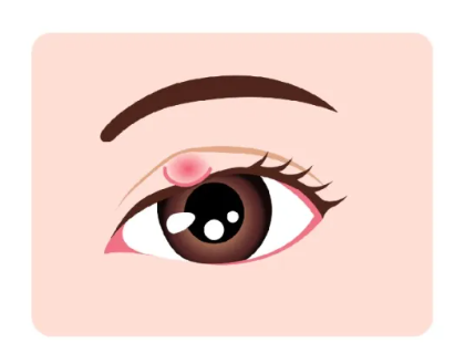
Ms Yu, aged 28, had been visiting various hospitals due to recurrent conjunctival haemorrhages and a gradually enlarging growth in the affected area. She recently visited the Cornea and Ocular Surface Department at Dongguan Aier Eye Hospital, where she informed the doctor that a growth had been developing in her right eye over the past six months and was gradually increasing in size.
Through a slit-lamp examination, Associate Chief Physician Ruan Yuanfei discovered that her right eye not only had a conjunctival naevus but also exhibited an abnormality whereby the right eyeball was noticeably more protruding than the left.
Based on clinical findings and further evaluation via magnetic resonance imaging (MRI), it was confirmed that a mass closely associated with the optic nerve had developed within the muscle cone located deep within the orbit and behind the eyeball. A comprehensive diagnosis was made of an orbital lymphangioma. “Intralesional haemorrhage can lead to sudden proptosis or enlargement of the eyeball, or even protrusion of the palpebral fissure, followed by haemorrhage in the eyelid or subconjunctival area.” As the tumour was relatively large, deeply situated and adjacent to vital neural structures, Associate Chief Physician Ruan Yuanfei recommended initially using corticosteroids to control and stabilise the condition whilst closely monitoring the patient, before formulating a subsequent treatment plan based on any changes.
In their early stages, these deep orbital tumours present with almost no noticeable symptoms. As they grow in size, they exert continuous pressure on the eyeball, causing it to protrude. They may also compress the optic nerve and the extraocular muscles, leading to vision loss and impaired eye movement; in severe cases, this can result in irreversible vision damage.
Many people assume that ‘protruding eyes are a sign of hyperthyroidism’. In fact, in clinical practice, protrusion is a common symptom of various orbital diseases; at best, it affects appearance, but at worst, it can threaten vision. Today, Dr Ruan Yuanfei will provide a systematic explanation of the common causes, warning signs, standardised examinations and diagnostic and treatment principles for eye protrusion, to help you identify the condition scientifically and seek timely medical attention.
A simple guide: How do you determine if your eyes are ‘protruding’?
In healthy adults, the normal protrusion of the eyeball is approximately 12–14 mm, with a difference of no more than 2 mm between the two eyes.
Simple self-check: Stand facing a mirror and look straight ahead. If one eye is noticeably bulging, there is asymmetry between the two eyes, or the cornea protrudes significantly beyond the orbital rim, this constitutes abnormal protrusion and you should seek an ophthalmological assessment as soon as possible.
Why do eyes protrude? An explanation of the four most common causes
Thyroid-associated ophthalmopathy: the most common cause of exophthalmos in adults
Thyroid-associated ophthalmopathy is the primary cause of unilateral or bilateral exophthalmos in adults; it can occur even when thyroid function is normal or reduced.
· Pathogenesis: Autoimmune abnormalities lead to oedema of the intraorbital soft tissues and hypertrophy of the extraocular muscles, which push the eyeball forward
· Typical symptoms: Proptosis in one or both eyes, retraction of the eyelids, dry eyes, photophobia, epiphora, diplopia, and impaired eye movement; in severe cases, compression of the optic nerve may result in reduced vision or even blindness
· Management: First, regulate thyroid function according to standard protocols; subsequently, the ophthalmologist will administer targeted treatments such as anti-inflammatory therapy, immunomodulation or orbital decompression
2. Orbital tumours: A high-risk cause of unilateral progressive exophthalmos
The orbital cavity is a confined space with intricate structures; tumours, whether benign or malignant, compress the eyeball and vital structures, making them a significant cause of unilateral progressive exophthalmos.
· Common types: cavernous haemangioma, lymphangioma, meningioma, lacrimal gland tumours, etc.
· Typical symptoms: Slow, unilateral protrusion of the eyeball; often painless in the early stages and easily overlooked; may be accompanied by reduced vision, double vision and restricted eye movement; eye pain and headaches may occur when nerves are compressed
· Note: Continued tumour growth significantly increases the difficulty and risk of surgery; once diagnosed, intervention should be sought as early as possible to avoid permanent damage to the optic nerve
3. Orbital inflammation: Acute exophthalmos with redness, swelling and pain
This is usually caused by swelling of the intraorbital tissues resulting from infectious or non-infectious inflammation, leading to sudden exophthalmos.
· Common types: Orbital cellulitis, inflammatory pseudotumour, etc.
· Typical symptoms: Protrusion of the eyeball accompanied by redness, swelling, warmth and pain; may be accompanied by fever and blurred vision; onset is usually acute
4. Other common causes
High myopia: The axial length of the eye increases, causing the eyeball to protrude; this is usually bilateral and symmetrical, with no redness, swelling or pain.
Vascular abnormalities / trauma: For example, a carotid-cavernous fistula may cause pulsatile exophthalmos, accompanied by a vascular murmur in the eye.
If you experience any of these warning signs, please seek medical attention immediately:
Protrusion of the eyeball is often an ‘early warning sign’ of orbital disease. You should seek medical attention as soon as possible if any of the following apply:
Progressive protrusion of one eye, or marked asymmetry between both eyes
Unexplained loss of vision, double vision, or visual field abnormalities
Eye pain, headache, eyelid swelling, or restricted eye movement
Inability to fully close the eyelids, dry eyes and sensitivity to light, or recurrent conjunctival haemorrhages
Unexplained “enlargement” of the eye, particularly if occurring on one side
Standardised examination and accurate diagnosis are prerequisites for treatment:
· Initial assessment: Slit-lamp examination, measurement of proptosis, visual acuity and fundus assessment
· Systemic assessment: Thyroid function tests and relevant antibody screening
· Accurate diagnosis: Orbital CT/MRI (provides clear visualisation of tumours, inflammation, and involvement of muscles and the optic nerve)
· Specialist consultation: Priority should be given to the lacrimal and orbital surgery department; complex cases require a multidisciplinary approach
Specialist Clinical Support:
Standardised Treatment for Complex Orbital Conditions
Orbital tumours are located deep within the eye socket and are situated close to nerves and blood vessels, making surgery highly complex and risky; they are considered among the most challenging procedures in ophthalmology. Since 2014, Dongguan Aier Eye Hospital has routinely performed intraorbital tumour surgery and has continued to specialise in complex eye conditions such as orbital and intraocular tumours.

In 2024, Director Liu Fei undertook specialised training at the University Hospital of Essen in Germany, where he systematically studied cutting-edge international techniques in the personalised diagnosis and treatment of ocular tumours, including radiotherapy, chemotherapy, laser therapy, cryotherapy and immunotherapy, and applied the principles of minimally invasive and precision medicine to clinical practice. The hospital has established a Lacrimal and Orbital Diseases Clinic, integrating imaging and multidisciplinary resources to create a standardised, personalised treatment system. It specialises in tackling complex eye conditions such as difficult-to-treat lacrimal disorders, orbital tumours and intraocular tumours. Through precise diagnosis, minimally invasive techniques and high-quality service, it provides high-standard diagnostic and treatment services for orbital diseases to patients in the Guangdong-Hong Kong-Macao Greater Bay Area.

2024年9月,刘斐院长远赴埃森大学医院研修

5月21日,Retina China 2026会议期间,刘斐院长、王虎院长与德国眼科学会前主席、德国埃森大学医院Bechrakis教授再度聚首,围绕眼内肿瘤诊疗前沿技术、学科建设、人才培养等核心内容展开深入交流。
A final reminder:
Protrusion of the eyeball is not merely a ‘cosmetic issue’, but an important warning sign regarding the health of the eye socket. Conditions such as thyroid-associated ophthalmopathy, orbital tumours, inflammation and vascular abnormalities can all lead to exophthalmos; in particular, unilateral progressive exophthalmos warrants vigilance for orbital tumours.
Early detection, early examination and early treatment are key to protecting your vision and reducing the risks associated with surgery. If you notice bulging eyes or experience any eye discomfort, seek prompt assessment at a reputable ophthalmology clinic, including orbital CT or MRI scans, to avoid permanent vision loss caused by delayed diagnosis and treatment.
PREV:Not all “lumps” on the eyelid are styes!
NEXT:...

咨询电话:
0769-22660023
医院地址:
东莞市东城区莞樟路石井路段171号-东莞爱尔眼科医院